A. sievert B. becquerel C. gray D. rem E. roentgen F. happy
2. The system unit for measuring the exposure dose of radiation is
A. gray B. curie C. rad D. X-ray E. becquerel F. coulomb / kg G. sievert H. rem
3. What fabrics are radio-resistant
A. bone B. lymphoid C. nervous D. cartilaginous E. myeloid F. intestinal epithelium
4. Among sulfur-containing radioprotectors, the RF Armed Forces use
A. cystamine B. cystaphos C. cystitone
5. The absorbed dose of radiation is -
A. The amount of radionuclides that entered the body by any means B. The amount of energy transferred by radiation to a substance per unit of its mass C. The radiation dose accumulated as a result of the absorption of radioactive isotopes D. The total charge of particles with electric charges of the same sign in the volume of air to mass air in this volume
6. The system unit of the absorbed radiation dose is
A. Sievert B. Gray C. Becquerel D. Rad E. X-ray F. rem G. Curie
7. Corpuscular types of ionizing radiation include
A. alpha radiation B. beta radiation C. gamma radiation D. X-rays
E. neutron radiation
8. What is called the result of the direct action of ionizing radiation?
A. a change in molecules that arise as a result of the absorption of energy by the molecules themselves B. a change in molecules caused by the products of radiolysis of water C. a change in molecules caused by the action of hydroperoxides
9. The processes occurring at the chemical stage under the action of ionizing radiation are
A. redistribution of absorbed energy within and between molecules B. formation of free radicals C. absorption of radiation energy D. repair and biological enhancement E. formation of ionized and excited molecules F. reactions of free radicals with each other and with intact biomolecules
10. Non-lethal reactions of cells to radiation include
A. radiation block of mitosis B. reproductive death C. interphase death D. impairment of specific functions E. mutations
11. Reproductive cell death is based on
A. genetically programmed mechanisms (apoptosis) B. damage to nuclear and mitochondrial membranes C. hyperactivation of poly-ADP-ribosylation processes D. chromosomal aberrations
12. The units of measurement of the exposure dose of radiation are
A. gray B. curie C. rad D. X-ray E. coulomb / kg F. Sievert G. rem
13. What types of radiation are rarely ionizing?
A. gamma radiation B. alpha radiation C. X-rays D. neutron radiation
14. The processes that make up the biological stage in the action of ionizing radiation are
A. redistribution of absorbed energy within and between molecules B. formation of free radicals C. absorption of radiation energy D. repair and biological enhancement of primary damage E. formation of ionized and excited atoms and molecules F. reactions between free radicals and their binding to biomolecules
15. Interphase form of cell death -
A. complete loss of the ability of cells to divide B. temporary loss of the ability of cells to divide C. deceleration of the process of cell division D. cell death without connection with the processes of cell division
16. What fabrics are highly radiosensitive?
A. bone B. lymphoid C. nervous D. cartilaginous E. myeloid
17. In experimental radiobiology, the most reliable quantitative characteristic of the effect of a radioprotector is
A. protection percentage B. protection factor C. radiation dose reduction factor (PDF)
18. The units of measurement of the absorbed dose of radiation 1 Gy and 1 rad are related as
A. 1 rad = 100Gy B. 1Gy = 1rad C. 1Gy = 100rad D. 1000rad = 1Gy
19. The radiation block of mitosis is -
A. complete loss of the ability of cells to divide
B. temporary loss of the ability of cells to divide
C. slowing down the process of cell division
D. death of dividing cells
20. The radiosensitivity of which cells does not correspond to the rule of Bergonier and Tribondo?
A. erythrocytes B. neurons C. lymphocytes D. basophils E. Clara cells
21. During the bone marrow form of acute radiation sickness, the following periods are distinguished
A. period of abortive fever B. period of recovery (resolution)
C. period of primary reaction to radiation (initial) D. lymphopenic
E. the peak period F. the period of imaginary well-being (latent)
G. period of primary devastation
22. To stop vomiting during the initial reaction to radiation, apply
A. cystamine B. dimetpramide C. athens D. dixaphene E. unitiol
23. What hematological changes are characteristic for the period of the primary reaction to radiation?
A. lymphopenia B. lymphocytosis C. neutrophilic leukocytosis D. erythropenia
24. The probable (stochastic) effects of human exposure include:
A. malignant tumors B. infertility C. radiation cataract
25. The main part of the radiation dose is received by the population of the Earth from
A. Natural sources of ionizing radiation
B. sources of ionizing radiation used in medicine
C. sources of ionizing radiation used in nuclear power
D. Fallout from nuclear explosions
26. The head of the medical service of the military unit is responsible for the organization of individual monitoring of radiation doses.
A. in the entire personnel of the medical service B. in the wounded and sick at the stages of medical evacuation C. in the entire personnel of the unit D. in the personnel during medical examinations
27. The most effective protection against gamma radiation is materials in which
A. heavy metals B. light metals C. hydrogen
28. In accordance with official documents, one-time exposure is called exposure, in which at least 80% of the dose an individual receives in no more than * days
29. The minimum dose of a single total external gamma-irradiation causing acute radiation sickness is estimated as * Gy
30. The minimum dose of a single total external gamma-irradiation causing acute radiation sickness in the bone marrow form is estimated as * Gy
31. The minimum dose of the total single external gamma-irradiation causing the intestinal form of acute radiation sickness is estimated as * Gy
32. The minimum dose of a single total external gamma irradiation causing the cerebral form of acute radiation sickness is estimated as * Gy
33. The development of acute radiation sickness of a mild degree can be expected with a total single uniform irradiation in doses from 1 to * Gy
34. The development of acute radiation sickness of moderate severity can be expected with a total single uniform external irradiation in doses from 2 to * Gy
35. The development of severe acute radiation sickness can be expected with a single uniform irradiation in doses from * to 6 Gy
36. The development of acute radiation sickness of an extremely severe degree can be expected with a total single uniform irradiation in doses exceeding * Gy
37. In acute radiation sickness of moderate severity in persons who have not taken antiemetics, vomiting is observed during the period of the initial reaction to radiation
A. single B. repeated C. multiple D. indomitable
A. in the first hours after irradiation B. 2 days after irradiation C. 7-9 days after irradiation D. at the end of the latent period
39. The probabilistic (stochastic) effect of human exposure includes:
A. malignant tumors B. infertility C. fetal malformations D. radiation cataract E. acute radiation sickness
40. The stochastic effects of irradiation are characterized by:
A. lack of a dose threshold B. proportionality of the severity of the effect to the dose
C. the probabilistic nature of the manifestation D. the presence of a dose threshold
41. The population of the world receives the main part of the dose of ionizing radiation from:
A. Natural sources B. NPP operation C. Nuclear weapons testing
D. use of sources of ionizing radiation in medicine
42. On the trail of a cloud of a nuclear explosion, servicemen receive the main radiation dose from:
A. external gamma radiation B. external beta radiation C. internal radiation
43. The head of the medical service of a military unit is responsible for organizing individual monitoring of radiation doses:
A. All personnel of the medical service B. Wounded and sick entering the stage of medical evacuation C. All personnel of the unit D. All personnel during a medical examination E. Command of the unit
44. Ionizing radiation includes:
A. ultrasonic radiation B. fast neutrons C. microwave radiation
D. "soft" X-rays
45. Indicate the units of measurement in the SI system for each of the listed types of radiation dose:
A. exposure B. absorbed C. equivalent a) Gy b) Sv c) C / kg
46. List three types of ionizing radiation in ascending order of their biological effectiveness for the human body under external irradiation:
A. beta radiation B. neutrons C. alpha radiation
47. Non-stochastic radiation effects are characterized by:
A. no dose threshold B. direct dependence of the severity of the effect on the dose
C. probabilistic character D. alternative character
48. The criteria for dividing the trace of a nuclear explosion cloud into zones of radioactive contamination include:
A. doses on the ground until the complete decay of the products of a nuclear explosion
B. radiation doses received by openly located personnel due to the action of penetrating radiation from a nuclear explosion
C. radiation doses received by openly located personnel due to the action of all radiation factors of a nuclear explosion
49. Victims with isolated radiation injuries will account for the largest share of sanitary losses in case of:
A. aerial nuclear explosion of ultra-small caliber ammunition
B. aerial nuclear explosion of medium caliber ammunition
C. underground nuclear explosion of medium caliber ammunition
D. ground-based nuclear explosion of an over-large caliber ammunition
50. The materials most effectively shielded from gamma radiation are materials in which predominate:
A. heavy metals B. light metals C. hydrogen D. carbon
51. The materials most effectively shielded from neutron radiation are materials in which predominate:
A. heavy metals B. light metals C. hydrogen D. nitrogen
52. Indicators characterizing the shielding ability of materials used for physical protection against ionizing radiation:
A. Linear energy transfer B. Half-attenuation layer C. Dose change factor D. Attenuation factor E. Linear ionization density
53. The absolute content of lymphocytes in the peripheral blood is a prognostic criterion for the severity of ARS from external irradiation:
A. in the first 1 day after irradiation B. 2-3 days after irradiation C. 8-9 days after irradiation D. at the end of the "latent" period E. in the first hours after irradiation
A. in the first hours after irradiation B. 1-2 days after irradiation C. 7-9 days after irradiation D. at the end of the "latent" period E. at the beginning of the peak period
55. Choose effective first aid measures when products of a nuclear explosion with contaminated food enter the body:
A. prescription of radioprotectors B. prescription of antiemetics C. gastric lavage D. prescription of saline laxatives E. colonic lavage
56. Choose effective first aid measures in case of contamination of eyes and exposed skin with products of a nuclear explosion:
A. appointment of radioprotectors B. appointment of antiemetics C. partial sanitization using IPP-11 D. application of a sterile cotton-gauze bandage to the infected skin area E. rinsing infected skin and eyes with clean water
57. The presence of widespread radiation erythema indicates radiation injury not less than:
A. mild ARS B. moderate ARS C. severe ARS
D. extremely severe ARS
58. Means of long-term maintenance of increased radioresistance of the body belong to the group
A. prophylactic antiradiation agents B. means of early pathogenetic therapy
C. means intended for the temporary preservation of the combat effectiveness of irradiated people
D. hospital therapy
59. The protective effect of radioprotectors is manifested in:
A. preserving the life of an irradiated person B. reducing the severity of radiation injury C. preventing the development of early transient incapacity for fighting D. relief of symptoms of a general primary reaction to radiation
60. The manifestation of the protective effect of radioprotectors is:
A. increase in the life span of an irradiated organism B. increase in the survival rate of irradiated people C. relief of symptoms of a general primary reaction to radiation D. prevention of symptoms of a general primary response to radiation
61. The use of radioprotectors is most effective in the following conditions:
A. pulsed irradiation B. irradiation with a dose rate above 0.02 Gy / min C. irradiation with a dose rate below 0.02 Gy / min D. prolonged irradiation E. fractionated irradiation
62. The mechanism of the radioprotective action of the drug B-190 is associated with:
A. interception of free radicals B. inhibition of the mitotic activity of bone marrow cells C. normalization of the physical state of excited molecules D. development of regional hypoxia E. oxygen effect
63. The factor of change in the dose of cystamine when administered to a person in the optimal radiation protective dose is estimated by the value:
A. 0.1 - 0.2 B. 0.2 - 1.2 C. 1.3 - 1.4 D. 1.4 - 1.7 E. 2.0 - 2.6
64. Specify radioprotectors from the group of imidazolines
A. indralin B. cystamine C. naphthyzine D. mexamine E. etiol
65. The representative of sulfur-containing radioprotectors is:
A. indralin B. diethylstilbestrol C. cystamine D. naphthyzine E. aminostigmine
66. Sulfur-containing radioprotectors include:
A. RS-1 B. EDS C. B-190 D. P-10M E. specimen C
67. Radioprotectors from the group of imidazolines include:
A. B-190 B. Specimen C C. RS-1 D. EDS E. P-10M
68. The means of long-term maintenance of increased radioresistance of the body are drugs belonging to the groups:
C. derivatives of imidazolines D. products derived from nucleic acids E. adaptogens of plant origin
69. Means of choice when working in conditions of low-intensity prolonged exposure are:
A. cystamine B. indralin C. riboxin D. tetrafolevite E. nicotinamide
70. Choose a product when working in conditions of prolonged exposure:
A. ginseng tincture B. ethaperazine C. indralin D. amitetravit E. naphthyzine
71. To prevent the manifestations of RPN-syndrome, you should use:
A. cystamine B. naphthyzine C. ethaperazine D. nicotinamide E. dixaphene
72. The mechanisms of radioprotective action of drugs for the prevention of RPN syndrome are:
A. modification of oxygen tension in tissues B. activation of the reticuloendothelial system C. inhibition of poly-ADP-ribosylation processes D. substrate provision of NAD-independent oxidation E. inactivation of free radicals
73. The mechanism of development of RPN-syndrome is associated with:
A. death of bone marrow stem cells B. stimulation of chemoreceptors in the trigger-zone of the medulla oblongata C. afferent impulses from mechano- and baroreceptors of the stomach
D. dysfunction of nerve cells
74. The procedure for using nicotinamide:
A. 6 tablets 30-60 minutes before irradiation B. 2 tablets 40-60 minutes before exposure
C. 1 tablet 20-30 minutes before irradiation D. 1 tablet at the first signs of injury
E. 10 tablets 1-24 hours before irradiation
75. The indication for the use of cystamine is the expected radiation dose:
A. 0.5 Gy and above B. 1 Gy and above C. 10 Gy and above D. 100 Gy and above E. 0.1 Gy and above
76. Choose a means to relieve symptoms of a primary reaction to radiation for an affected person with severe ARS:
A. naphthyzine B. indralin C. latran D. dimetpromide E. nicotinamide
77. Choose means to relieve symptoms of a general primary reaction to radiation:
A. cystamine B. dixafen C. zofran D. nicotinamide
Any substances, living organisms and their tissues.
Collegiate YouTube
1 / 5
✪ How exactly does radiation kill?
✪ More about radiation
✪ Radiation sickness
✪ Alpha, beta and gamma radiation | Physics Grade 11 # 47 | Info lesson
✪ Dose rate of gamma radiation
Subtitles
Hello everyone! Dmitry Pobedinsky is with you and I am glad to welcome you to the QWERTY Channel! comrades, let's remember school classes in Warsaw there was a lot of something in Prague separate explosions in the bar and bombs the shelter ended up letting go I don’t remember the details one year of them definitely radiation is dangerous and sometimes even fatal, but I wonder how exactly radiation beats just from the outside everything is clear the bullet is a fool or that they make a hole in their business, I start chemical reactions and communicators threaten them, but also the action of exactly how it affects a person, let's first remember what we already imagine to be reduced to a size 10 thousand times smaller than an atom then we can see then where do the main types of radiation come from the atomic nucleus, as we remember, it consists of protons and there are no mouths, and I know for some alimony it can be configured cop, roughly speaking, not quite the way it becomes unstable in them there is extra energy and in which they strive to get rid of and this can be done in several ways by throwing out a small piece of d va protons two neutrons are they to be cleaned in ugra a neutron can turn into a proton and vice versa, then this electron antirecord flies into this particle, its twin only with the opposite sign, and finally the nucleus can simply throw out if, when children, anticipating an electromagnetic wave, this one, like ultraviolet light, led this prime legs can also cleanse the bowels of the earth can emit neutrons protons broke into pieces besides, radiation particles can fly from space appears in accelerators and other devices, but despite the differences in the origin and restructuring of any types of radiation, the most important thing on the body is that this particle flux is at the rate of and energy, the impact of radiation on a person who looks like a snowball, everything starts small, but then the consequences grow and grow until they lead to irreversible changes, several stations can be distinguished so the radiation particles of the face are faster than any path so quickly that they knock out electrons from the tents, the electrode is negative, respectively, the act of receiving the loss becomes positive ions, that's all that radiation does, but the flow of free electrons and they are isolated atom almost immediately participate in complex reaction chains in which a chemically active molecule can be formed, including the so-called free radicals well, for example, the water of which a person consists of 80 percent of us under the influence of radiation breaks down into two radicals, as much as its free radicals actively react with important biological molecules dorenko beat Chirac chambers with experiments, as a result of which the molecule is damaged from them, toxins are often formed, the normal metabolism of the cell is disrupted by its functioning in general, and after a while, it dies, but even if the cell is strong in the spirit of the hero, it holds on to the last, it is still doomed, because due to DNA damage and gene mutations, normal cell division is impossible, this is perhaps the most dangerous There is a lot of radiation with a large dose of radiation, the affected cells are very much and whole can refuse only to find systems that are most susceptible to radiation, tissues in which active cell division is taking place, for example, the bone marrow in which blood is being processed or a consequence of the stomach that is expected by acid and should be actively regenerated, summing up, we can say radiation acts on the smallest scale in the structure of the human body, it is as if they fired at the exit of the fortress wall unprepared by shells and small small bullets so that the damage can be easily repaired, however if the field is huge, then the damage will be repaired and in the hands the wall will eventually become fragile and sooner or later will fall apart but you will never be able to hide from radiation with him it follows us everywhere in almost every substance there is a small fraction of unstable isotopes so there is a little radioactive around us in seoul computers video cameras apples ban now, but even people in a person, for example, every second there are several thousand radioactive decays, it's another matter and the radiation intensity of course, the radiation of ordinary objects is very weak, well, and safe background radiation in general could be the driving force of the revolution, because perhaps it was thanks to her that the genes mutated so that we ended up like this it is cool to understand how to protect yourself from an excessive dose of radiation, the radiation attack will be easily saved by cardboard sheets, otherwise you can hide behind glass, but gamma radiation penetrates everything worse than an X-ray, so you can escape from it only for a thick layer of lead; another thing if the source gets in breathe out radioactive dust into your body or eat something, then all types of radiation will act on the body from the inside and the consequences will be much more serious in terms of radiation there is no smell, no color or
Exposure dose
The main characteristic of the interaction of ionizing radiation with a medium is the ionization effect. In the initial period of the development of radiation dosimetry, most often it was necessary to deal with X-rays propagating in the air. Therefore, the degree of air ionization was used as a quantitative measure of the radiation field. A quantitative measure based on the amount of ionization of dry air at normal atmospheric pressure, which is quite easy to measure, is called exposure dose.
The exposure dose determines the ionizing capacity of X-rays and gamma rays and expresses the radiation energy converted into kinetic energy of charged particles per unit mass of atmospheric air. The exposure dose is the ratio of the total charge of all ions of the same sign in an elementary volume of air to the mass of air in this volume.
| Type of radiation | Coefficient, Sv / Gy |
|---|---|
| X-ray and γ-radiation | 1 |
| β-radiation (electrons, positrons) | 1 |
| Neutrons with energies less than 20 keV | 3 |
| Neutrons with energies 0.1-10 MeV | 10 |
| Protons with energies less than 10 MeV | 10 |
| α-radiation with energies less than 10 MeV | 20 |
| Heavy recoil kernels | 20 |
Effective dose
Effective dose (E) is a value used as a measure of the risk of long-term effects of irradiation of the entire human body and its individual organs and tissues, taking into account their radiosensitivity. It represents the sum of the products of the equivalent dose in organs and tissues by the corresponding weighting factors.
The value of the radiation risk coefficient for individual organs
| Organs, tissues | Coefficient |
| Gonads (sex glands) | 0,2 |
| Red bone marrow | 0,12 |
| Colon | 0,12 |
| Stomach | 0,12 |
| Lungs | 0,12 |
| Bladder | 0,05 |
| Liver | 0,05 |
| Esophagus | 0,05 |
| Thyroid | 0,05 |
| Leather | 0,01 |
| Bone surface cells | 0,01 |
| Brain | 0,05 |
| Other fabrics | 0,05 |
Weighted coefficients are established empirically and calculated in such a way that their sum for the whole organism is one. The units of measurement for the effective dose are the same as those for the equivalent dose. It is also measured in sievert or rem.
Fixed effective equivalent dose(CEDE - the committed effective dose equivalent) is an estimate of radiation doses to a person, as a result of inhalation or consumption of a certain amount of a radioactive substance. CEDE is expressed in rem or sievert (Sv) and takes into account the radiosensitivity of various organs and the time during which the substance remains in the body (up to the whole life). Depending on the situation, CEDE may also relate to the radiation dose to a specific organ rather than the whole body.
Effective and equivalent dose- these are standardized values, that is, values that are a measure of damage (harm) from the impact of ionizing radiation on a person. Unfortunately, they cannot be directly measured. Therefore, operational dosimetric quantities have been introduced into practice, which are uniquely determined through the physical characteristics of the radiation field at a point, as close as possible to the standardized ones. The main operating quantity is the ambient dose equivalent (synonyms - ambient dose equivalent, ambient dose).
Ambient dose equivalentН * (d) - dose equivalent, which was created in a ball phantom ICRU (International Commission on Radiation Units) at a depth d (mm) from the surface in diameter parallel to the direction of radiation, in a radiation field identical to that considered in composition, fluence and energy distribution, but unidirectional and homogeneous, that is, the ambient dose equivalent H * (d) is the dose that a person would receive if he were at the place where the measurement is carried out. The unit of the ambient dose equivalent is the sievert (Sv).
Group doses
By calculating the individual effective doses received by individuals, one can come to the collective dose - the sum of the individual effective doses in a given group of people over a given period of time. The collective dose can be calculated for the population In addition, the following doses are distinguished:
- commitment - the expected dose, half a century dose. Used in radiation protection and hygiene when calculating absorbed, equivalent and effective doses from incorporated radionuclides; has the dimension of the corresponding dose.
- collective - a calculated value introduced to characterize the effects or damage to health from irradiation of a group of people; unit - Sievert (Sv). The collective dose is defined as the sum of the products of average doses by the number of people in dose intervals. A collective dose can accumulate over a long time, not even one generation, but covering subsequent generations.
- threshold - the dose below which the manifestations of this radiation effect are not observed.
- maximum permissible doses (PDD) - the highest values of the individual equivalent dose for a calendar year, at which uniform irradiation for 50 years cannot cause adverse changes in the state of health detected by modern methods (NRB-99)
- avoidable - the predicted dose due to a radiation accident, which can be prevented by protective measures.
- doubling - a dose that doubles (or 100%) the level of spontaneous mutations. The doubling dose is inversely proportional to the relative mutational risk. According to the currently available data, the value of the doubling dose for acute exposure is on average 2 Sv), and for chronic exposure it is about 4 Sv.
- biological dose of gamma-neutron radiation - a dose of gamma-radiation, which is equally effective in damaging the body, taken as a standard one. Equal to the physical dose of the given radiation multiplied by the quality factor.
- minimum lethal - the minimum dose of radiation that causes the death of all irradiated objects.
Dose rate
Dose rate(radiation intensity) - the increment of the corresponding dose under the influence of the given radiation per unit of time. Has the dimension of the corresponding dose (absorbed, exposure, etc.), divided by a unit of time. The use of various special units is allowed (for example, Sv / h, rem / min, mSv / year, etc.).
Units summary table
| Physical quantity | Non-system unit | SI unit | Transition from a non-SI unit to a SI unit |
|---|---|---|---|
| Nuclide activity in a radioactive source | Curie (Ki) | Becquerel (Bq) | 1Ci = 3.7⋅10 10 Bq |
| Exposure dose | X-ray (R) | Pendant / kilogram (C / kg) | 1P = 2.58⋅10 −4 C / kg |
| Absorbed dose | Glad (glad) | Gray (J / kg) | 1rad = 0.01 Gy |
| Equivalent dose | Rem (rem) | Sievert (Sv) | 1 rem = 0.01 Sv |
| Exposure dose rate | X-ray / second (R / s) | Pendant / kilogram per second (C / kg * s) | 1Р / s = 2.58⋅10 −4 C / kg * s |
| Absorbed dose rate | Rad / second (Rad / s) | Gray / second (Gy / s) | 1 rad / s = 0.01 Gy / s |
| Equivalent dose rate | Rem / second (rem / s) | Sievert / second (Sv / s) | 1 rem / s = 0.01 Sv / s |
| Integral dose | Rad-gram (Rad-g) | Gray-kilogram (Gr-kg) | 1rad-g = 10 −5 Gy-kg |
1. Dosimetry. Radiation doses. Dose rate.
2. Biological effects of radiation doses. Limiting doses.
3. Dosimetric devices. Ionizing radiation detectors.
4. Methods of protection against ionizing radiation.
5. Basic concepts and formulas.
6. Tasks.
34.1. Dosimetry. Radiation doses. Dose rate
The need for a quantitative assessment of the effect of ionizing radiation on various substances of animate and inanimate nature led to the emergence of dosimetry.
Dosimetry - the section of nuclear physics and measuring technology, which studies the quantities characterizing the effect of ionizing radiation on substances, as well as methods and instruments for their measurement.
The processes of interaction of radiation with tissues proceed differently for different types of radiation and depend on the type of tissue. But in all cases, there is a transformation of radiation energy into other types of energy. As a result, part of the radiation energy is absorbed by the substance. Absorbed energy- the root cause of all subsequent processes, which ultimately lead to biological changes in a living organism. Quantitatively, the effect of ionizing radiation (regardless of its nature) is estimated by the energy transferred to the substance. For this, a special value is used - radiation dose(dose - portion).
Absorbed dose
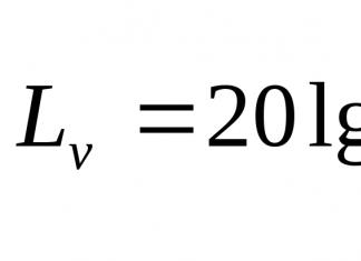
Absorbed dose(D) is a quantity equal to the ratio of energyΔ Ε, transferred to the element of the irradiated substance, to the massΔ m of this element:
In SI, the unit of absorbed dose is gray (Gr), in honor of the English physicist-radiobiologist Louis Harold Gray.
1 Gy - it is the absorbed dose of ionizing radiation of any kind, at which 1 J of radiation energy is absorbed in 1 kg of the substance mass.
In practical dosimetry, an off-system unit of absorbed dose is usually used - glad(1 glad= 10 -2 Gr).
Equivalent dose
The quantity absorbed dose takes into account only the energy transferred to the irradiated object, but does not take into account the "quality of radiation". Concept radiation quality characterizes the ability of a given type of radiation to produce various radiation effects. To assess the quality of radiation, the parameter is introduced - quality factor. It is a regulated value, its values are determined by special commissions and are included in international standards designed to control radiation hazard.
Quality factor(K) shows how many times the biological effect of this type of radiation is greater than the effect of photon radiation, at the same absorbed dose.
Quality factor is a dimensionless quantity. Its values for some types of radiation are given in table. 34.1.
Table 34.1. Quality factor values
 Equivalent dose(H) is equal to the absorbed dose multiplied by the quality factor for the given type of radiation:
In SI, the unit of equivalent dose is called sievert (Sv) - in honor of the Swedish specialist in the field of dosimetry and radiation safety Rolf Maximilian Sievert. As well as sievert a non-systemic unit of equivalent dose is also used - rem(biological equivalent of X-ray): 1 rem= 10 -2 Sv.
If the body is exposed several types of radiation, then their equivalent doses (H i) summarize:
Effective dose
With a single general irradiation of the body, different organs and tissues have different sensitivity to the action of radiation. So, with the same equivalent dose the risk of genetic damage is most likely with irradiation of the reproductive organs. The risk of lung cancer when exposed to α-radiation from radon under equal exposure conditions is higher than the risk of skin cancer, etc. Therefore, it is clear that the radiation doses of individual elements of living systems should be calculated taking into account their radiosensitivity. To do this, use the weight coefficients b T (T is the index of the organ or tissue), given in table. 34.2.
Table 34.2. The values of the weight coefficients of organs and tissues when calculating the effective dose
 The end of the table. 34.2
 Effective dose(H eff) is a value used as a measure of the risk of long-term effects of irradiation of the entire human body, taking into account the radiosensitivity of its individual organs and tissues.
Effective dose is equal to the sum of the products of equivalent doses in organs and tissues by the corresponding weight coefficients:
 The summation is carried out over all the tissues listed in table. 34.2. Effective doses, as well as equivalent ones, are measured in rem and sievert.
Exposure dose
The absorbed and associated equivalent radiation dose is characterized by energetic action radioactive radiation. As a characteristic ionizing action radiation use another quantity called exposure dose. Exposure dose is a measure of air ionization by X-rays and γ-rays.
Exposure dose(X) is equal to the charge of all positive ions generated by radiation per unit mass of air under normal conditions.
In SI, the unit of exposure dose is pendant per kilogram (C / kg). Pendant - this is a very large charge. Therefore, in practice, they use a non-systemic unit of the exposure dose, which is called x-ray(P), 1 R= 2.58x10 -4 Cl / kg. At an exposure dose of 1 R as a result of ionization in 1 cm 3 of dry air under normal conditions, 2.08x10 9 pairs of ions are formed.
The relationship between absorbed and exposure doses is expressed by the ratio
where f is some conversion factor depending on the irradiated substance and the radiation wavelength. In addition, the f value depends on the dose units used. F-values for units glad and x-ray are given in table. 34.3.
Table 34.3. Conversion factor values from x-ray v glad
 In soft tissues f ≈ 1, therefore the absorbed dose of radiation in glad is numerically equal to the corresponding exposure dose in x-rays. This leads to the convenience of using non-systemic units glad and R.
Relationship between different doses are expressed by the following formulas:
Dose rate
Dose rate(N) is a value that determines the dose received by an object per unit of time.
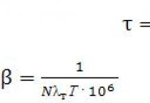
With uniform exposure to radiation dose rate is equal to the ratio of the dose to the time t, during which the ionizing radiation acted:
where κ γ is the gamma constant characteristic of the given radioactive preparation.
Table 34.4 shows the relationship between dose units.
Table 34.4. Relationship between dose units
 34.2. Biological effects of radiation doses. Limiting doses
The biological effect of radiation with different equivalent doses is indicated in table. 34.5.
Table 34.5. Biological action of single effective doses
 Limiting doses
Radiation safety standards establish dose limits(PD) of radiation, compliance with which ensures the absence of clinically detectable biological effects of radiation.
Limiting dose- the value of the annual effective the dose of technogenic radiation, which should not be exceeded under normal operating conditions.
The values of the limiting doses are different for staff and population. Personnel are persons working with man-made sources of radiation (group A) and under the working conditions in the area of their impact (group B). For group B, all dose limits are set four times lower than for group A.
For the population, the dose limits are 10-20 times less than for group A. The PD values are given in table. 34.6.
Table 34.6. Main dose limits
 Natural (natural) radiation background created by natural radioactive sources: cosmic rays (0.25 mSv / year); radioactivity of the bowels (0.52 mSv / year); radioactivity of food (0.2 mSv / year).
Effective dose up to 2 mSv / year(10-20 μR / h), obtained through natural background radiation, considered normal. As in the case of man-made irradiation, the level of irradiation more than 5 is considered high. mSv / year.
There are places on the globe where the natural background is 13 mSv / year.
34.3. Dosimetric devices. Ionizing radiation detectors
Dosimeters- measuring devices doses ionizing radiation or dose related quantities. The dosimeter contains detector radiation and measuring device, which is graduated in units of dose or rate.
Detectors- devices that register various types of ionizing radiation. The work of detectors is based on the use of those processes that cause registered particles in them. There are 3 groups of detectors:
1) integral detectors,
2) counters,
3) track detectors.
Integral detectors
These devices provide information on the total flow of ionizing radiation.
1. Photodosimeter. The simplest integral detector is an opaque X-ray film cassette. A photodosimeter is an individual integral meter that is supplied to persons who come into contact with radiation. The film appears after a certain period of time. The radiation dose can be determined by the degree of its blackening. Detectors of this type can measure doses from 0.1 to 15 R.
2. Ionization chamber. This is a device for registering ionizing particles by measuring the amount of ionization (the number of ion pairs) produced by these particles in a gas. The simplest ionization chamber consists of two electrodes placed in a gas-filled volume (Fig. 34.1).
A constant voltage is applied to the electrodes. Particles entering the space between the electrodes ionize the gas, and a current is generated in the circuit. The current strength is proportional to the number of ions formed, i.e. exposure dose rate. The electronic integrating device determines the X dose itself.
 Rice. 34.1. Ionization chamber
Counters
These devices are designed to count the number of particles of ionizing radiation passing through working volume or falling on work surface.
1. Figure 34.2 shows a diagram of a gas-discharge Geiger-Muller counter, the principle of operation of which is based on the formation of an electric pulse discharge in a gas-filled chamber when a separate ionizing particle hits.
 Rice. 34.2. Geiger-Muller counter circuit
The counter is a glass tube with a metal layer (cathode) sprayed onto its lateral surface. A thin wire (anode) is passed inside the tube. The gas pressure inside the tube is 100-200 mm Hg. A high voltage of the order of hundreds of volts is created between the cathode and anode. When an ionizing particle enters the counter, free electrons are formed in the gas, which move to the anode. The field strength is high near the thin filament of the anode. Electrons near the filament are accelerated so much that they begin to ionize the gas. As a result, a discharge occurs and a current flows through the circuit. The self-sustained discharge must be extinguished, otherwise the counter will not react to the next particle. There is a significant voltage drop across the high-resistance R connected in the circuit. The counter voltage decreases and the discharge stops. Also, a substance is introduced into the composition of the gas, corresponding to the fastest extinguishing of the discharge.
2. An improved version of the Geiger-Muller counter is proportional counter, in which the amplitude of the current pulse is proportional to the energy released in its volume by the detected particle. Such a counter determines absorbed dose radiation.
3. Action is based on another physical principle scintillation counters. Scintillations occur in some substances under the influence of ionizing radiation, i.e. flashes, the number of which is counted using a photomultiplier tube.
Track detectors
Detectors of this type are used in scientific research. V track detectors the passage of a charged particle is recorded in the form of a spatial picture of the track (track) of this particle; the painting can be photographed or registered with electronic devices.
A common type of track detector is Wilson chamber. The observed particle passes through a volume filled with oversaturated steam, and ionizes its molecules. Vapor condensation begins on the formed ions, as a result of which the particle track becomes visible. The camera is placed in a magnetic field that bends the trajectories of charged particles. The curvature of the track can be used to determine the mass of the particle.
34.4. Methods of protection against ionizing radiation
Protection against the negative effects of radiation and some ways to reduce the radiation dose are listed below. There are three types of protection: protection by time, distance and material.
Time and distance protection
For a point source, the exposure dose is determined by the ratio
from which it can be seen that it is directly proportional to time and inversely proportional to the square of the distance to the source.
A natural conclusion follows from this: to reduce the damaging radiation effect, it is necessary to stay as far as possible from the radiation source and, if possible, for less time.
Material protection
If the distance to the radiation source and the exposure time cannot be kept within safe limits, then it is necessary to protect the body with the material. This method of protection is based on the fact that different substances absorb in different ways all kinds of ionizing radiation that fall on them. Depending on the type of radiation, protective screens made of various materials are used:
alpha particles- paper, a layer of air several centimeters thick;
beta particles- glass several centimeters thick, aluminum plates;
X-ray and gamma radiation- concrete 1.5-2 m thick, lead (these emissions are attenuated in the substance according to an exponential law; a large thickness of the shielding layer is needed; in X-ray rooms, a lead-lined rubber apron is often used);
neutron flux- slows down in hydrogen-containing substances, such as water.
For personal protection of the respiratory system from radioactive dust are used respirators.
In emergency situations associated with nuclear disasters, you can take advantage of the protective properties of residential buildings. So, in the basements of wooden houses, the external radiation dose is reduced by 2-7 times, and in the basements of stone houses - by 40-100 times (Fig. 34.3).
When radioactive contamination of the area is controlled activity one square kilometer, and if food is contaminated, their specific activity. As an example, it can be indicated that when an area is infected with more than 40 Ci / km 2, the inhabitants are completely resettled. Milk with a specific activity of 2x10 11 Ci / l and more should not be consumed.
 Rice. 34.3. Shielding properties of stone and wooden houses for external γ-radiation
34.5. Basic concepts and formulas
 Table continuation
 End of the table
 34.6. Tasks
1. The study of radiation cataracts in rabbits showed that under the influence of γ - radiation from cataracts develops at a dose of D 1 = 200 rad. Under the action of fast neutrons (accelerator halls), cataract occurs at a dose of D 2 = 20 rad. Determine the quality factor for fast neutrons.
 2.
How many degrees will the temperature of a phantom (model of a human body) with a mass of 70 kg increase with a dose of γ-radiation X = 600 R? Specific heat capacity of the phantom c = 4.2x10 3 J / kg. Consider that all the energy received is used for heating.
 3.
A person weighing 60 kg was exposed to γ-radiation for 6 hours, the power of which was 30 μR / hour. Assuming that the main absorbing element is soft tissue, find the exposure, absorbed and equivalent radiation dose. Find the absorbed radiation energy in SI units.
 4.
It is known that a single lethal exposure dose for humans is 400 R(50% mortality). Express this dose in all other units.
5. In a tissue with a mass of m = 10 g, 10 9 α-particles with an energy of E = 5 MeV are absorbed. Find the equivalent dose. The quality factor for α-particles is K = 20.
 6.
Exposure dose rate γ
-radiation at a distance r = 0.1 m from a point source is N r = 3 R / hour. Determine the minimum distance from the source at which you can work 6 hours daily without protection. PD = 20 mSv / year. Absorption γ
- disregard air emissions.
Solution(accurate alignment of units required) Radiation Safety Standards equivalent dose, obtained for a year of operation is H = 20 mSv. Quality factor for γ -radiation K = 1.
 Applications
Fundamental physical constants

 Multipliers and prefixes for the formation of decimal multiples and sub-multiples and their designations
The result of radiation exposure depends on a number of factors: the amount of radioactivity in the external environment and inside the body, the type of radiation and its energy during the decay of the nuclei of radioactive isotopes, the accumulation of radioactive substances in the body and their excretion, etc. dissipated mass of substance. As a result of the interaction of radioactive radiation with the environment, including biological objects, a certain amount of radiation energy is transferred to it, which is spent on the processes of ionization and excitation of atoms and molecules of the medium. Part of the radiation passes through the medium freely, without absorption, without affecting it. Therefore, there is a direct relationship between the effect of radiation and the amount of absorbed energy. This determines the radiation dose.
Dose is understood as the measure of the effect of ionizing radiation in a certain environment.
Dose- the amount of radiation energy transferred to the substance and calculated per unit mass or volume of the substance.
With an increase in the exposure time of the object, the dose increases.
To measure the amount of absorbed energy, it is necessary to count the number of ion pairs formed under the action of ionizing radiation. In this regard, for the quantitative characteristics of X-ray and gamma radiation acting on an object, the concept was introduced "Exposure dose".
Exposure dose (X)- dose, which characterizes the ionization capacity of X-ray or gamma radiation (photon radiation) in air at a photon energy of no more than 3 MeV. It is also called physical.
The exposure dose is the ratio of the total charge dQ of all ions of the same sign created in the air, when all electrons and positrons released by photons in an elementary volume of air with mass dm completely stopped in air, to the mass of air in the specified volume:
The exposure dose is used to assess the radiation situation on the ground, in the work or living room, caused by the action of X-ray or gamma radiation, as well as to determine the degree of protective properties of screen materials.
The unit of exposure dose in the International System of Units (SI) is a pendant per kilogram (C / kg).
Pendant per kilogram This is such an exposure dose of X-ray or gamma radiation, at which the conjugated corpuscular emission (all electrons and positrons released by photons) in a volume of air weighing 1 kg produces ions carrying an electric charge of one coulomb (C) of each sign (+ and -).
From 1.01.1990, off-system units expressing dose and activity were to be withdrawn from use (R, Rad, Baer, Ki, etc.). However, they are still in use, which is explained, in particular, by the use in practice of a fleet of dosimetric and radiometric instruments with calibration of recording devices in non-systemic units.
The non-systemic unit of measurement of the exposure dose is X-ray (R). This unit has been in circulation since 1928.
X-ray- exposure dose of X-ray or gamma radiation, at which 2.08 · 10 9 pairs of ions are formed in 1 cm 3 (0.001293 g) of air under normal conditions (temperature 0 ° C and pressure 760 mm Hg). Or x-ray- exposure dose of X-ray or gamma radiation, at which the conjugate corpuscular emission in 1 cm 3 of air under normal conditions creates ions that carry a charge in one electrostatic unit of electricity of each sign.
1 P = 2.58 · 10 -4 C / kg; 1 C / kg = 3.88 · 10 3 R
An exposure dose of 1 X-ray is created by gamma radiation from a radium source with an activity of 1 Ci at a distance of 1 meter in 1 hour.
Derivative X-ray units: kilo-roentgen (1 kR = 10 3 R), millirent-gene (1 mR = 10 -3 R), micro-roentgen (1 μR = 10 -6 R).
For corpuscular ionizing radiation (alpha and beta particles, neutrons), a non-systemic unit was proposed - the physical equivalent of an X-ray (fer), in which the same number of ion pairs are formed in the air as with an exposure dose of X-ray or gamma radiation in 1 R. Unit Fair has not received any practical application and is currently not used. To characterize the radiation fields, it is better to use the particle flux density (including photons) and the radiation intensity (energy flux density).
The exposure dose is unacceptable to corpuscular types of radiation (alpha and beta particles, etc.), is limited to the energy range of quanta up to 3 MeV and reflects only a measure of the amount of photon radiation. It does not reflect the amount of radiation energy absorbed by the irradiated object. At the same time, it is very important to know the amount of radiation energy absorbed by the object for assessing the radiation effect. To determine the measure of the absorbed energy of any type of radiation in a medium, the concept was introduced "Absorbed dose". By the magnitude of the absorbed dose, knowing the atomic composition of the substance, the radiation energy, it is possible to calculate the absorbed dose of X-ray and gamma radiation in any substance. The energy equivalent of X-rays is 88 erg / g (the energy spent on the formation of 2.08 · 10 9 pairs of ions).
Absorbed dose (D)- the value of the energy of ionizing radiation transferred to the substance:
where de is the average energy transferred by ionizing radiation to a substance located in an elementary volume, dm is the mass of a substance in this volume.
Or absorbed dose- the amount of energy of any kind of ionizing radiation absorbed in a particular organ or tissue and calculated per unit mass.
If we denote the energy that falls on the object by the value E, and the energy passed through the object - E 1, then ∆E will be the absorbed energy:
∆Е = Е - Е 1.
Instead of the term "absorbed radiation dose", the abbreviated form "radiation dose" may be used.
The unit of measure for absorbed dose in the International System of Units is joule per kilogram (J / kg).
Joule per kilogram- such a unit of absorbed dose, at which 1 joule of energy is absorbed by any type of ionizing radiation in 1 kg of the mass of the irradiated substance.
This unit is called gray (Gr) in a different way.
Gray - a unit, like the off-system unit X-ray, is eponymous, that is, it is formed on behalf of the scientist. Louis Harold Gray is an English radiobiologist who worked on the relationship between the physical and biological effects of radiation and made a great contribution to the development of radiation dosimetry.
Gray is equal to the absorbed dose of radiation at which a substance with a mass of 1 kg is transferred an ionizing radiation energy equal to 1 J (1 Gy = 1 J / kg).
Derived units from gray are also used: μGy, mGy, etc.
Since 1953, a non-systemic unit of absorbed dose was introduced - rad (from the English radiation absorbed dose - absorbed dose of radiation), which is still widely used in practice today.
Glad- the absorbed dose of any kind of ionizing radiation, at which a radiation energy equal to 100 erg is absorbed in 1 g of the substance.
1 rad = 100 erg / g = 10 -2 J / kg; 100 rad = 1 Gr.
Fractional and multiple units of rad are used: kilorad (1 krad = 10 3 rad), millirad (1 mrad = 10 -3 rad), microrad (1 mrad = 10 -6 rad).
To calculate the absorbed dose, use the formula:
where D is the absorbed dose, X is the exposure dose, F is the transition coefficient, established empirically on the phantom (for water and soft tissue, F is 0.93 or ≈ 1).
In air, a radiation dose of 1 X-ray is energetically equivalent to 88 erg / g, the absorbed dose from the definition is 100 erg / g, therefore, the absorbed dose in air will be 0.88 rad (88: 100 = 0.88).
Under conditions of radial equilibrium, in which the sum of energies of charged particles leaving the considered volume corresponds to the sum of energies of charged particles entering this volume, it is possible to establish the energy equivalent of the exposure dose.
The exposure dose in air X = 1 P corresponds to the absorbed dose D = 0.873 rad, and 1 C / kg = 33.85 Gy. In biological tissue: 1 P corresponds to 0.96 rad and 1 C / kg corresponds to 33.85 Gy. Thus, with a small error (up to 5%) under uniform photon irradiation, the absorbed dose in biological tissue coincides with the exposure dose measured in X-rays.
When living organisms are irradiated, various biological effects arise, the difference between which at the same absorbed dose is explained by the degree of danger for the organism of different types of radiation.
It is customary to compare the biological effects caused by any ionizing radiation with the effects of photon, that is, X-ray and gamma radiation, as well as the spatial distribution of the absorbed energy in the irradiated object. At the same absorbed dose, alpha radiation is much more dangerous than beta or gamma radiation. To take this phenomenon into account, the concept was introduced "Equivalent dose".
Equivalent dose (N) Is the absorbed dose in an organ or tissue multiplied by the appropriate weighting factor for a given type of radiation (W R):
H TR = D TR W R,
where D TR is the average absorbed dose in an organ or tissue T, W R is a weighting factor for radiation R.
When an object is exposed to various types of radiation with different weighting factors, the equivalent dose is determined as the sum of the equivalent doses for these types of radiation.
The equivalent dose is the main quantity that determines the level of radiation hazard during chronic exposure of humans and animals in low doses.
In the international system of units (SI), a sievert (Sv) is taken as a unit of equivalent dose. The sievert unit is intended for use in the field of radiation safety only.
This unit of measure of equivalent dose was named after the Swedish scientist Rolf Sievert, who was engaged in research in the field of dosimetry and radiation safety.
Sievert is an equivalent dose of any kind of radiation absorbed by 1 kg of biological tissue and creating the same biological effect as an absorbed dose of 1 Gy of photon radiation.
The non-systemic unit of measurement of the equivalent dose is rem (abbreviation - biological equivalent of X-ray).
Rem is an equivalent dose of any type of ionizing radiation, at which the same biological effect is created in biological tissue as with a dose of X-ray or gamma radiation of 1 roentgen.
1 rem = 1 · 10 -2 J / kg;
100 rem = 1 Sv.
Weighting factors for individual types of radiation when calculating the equivalent dose (W R)- the absorbed dose multipliers used in radiation protection, taking into account the relative efficiency of various types of radiation in inducing biological effects. Previously, a quality factor (Q) or relative biological effectiveness (RBE) was used for this purpose.
The radiation quality factor is designed to take into account the influence of the microdistribution of absorbed energy on the degree of manifestation of a harmful biological effect and is selected on the basis of the available RBE coefficient values.
The RBE coefficient, or (Q), shows how many times the effectiveness of the biological action of a given type of radiation is greater than that of X-ray or gamma radiation at the same absorbed dose in tissues. The higher the specific ionization, the higher the RBE coefficient, or (Q).
Weighting factors (W R) for individual types of radiation:
Photons of any energy (X-ray or gamma radiation) ... ... 1
Electrons (beta particles) ………………………………………… ..1
Alpha particles, fission fragments, heavy nuclei ……………. …… 20
There are also the following types of doses: effective, effective expected from internal irradiation, effective collective and effective annual.
Effective dose (E)- a value used as a measure of the risk of the long-term effects of irradiation of the whole body and its individual organs, taking into account their radiosensitivity. It represents the sum of the products of the equivalent dose in the organ Н tТ by the corresponding weighting factor for the given organ or tissue:
Е = ∑W Т Н tТ,
where Н tТ is the equivalent dose in the tissue during the time t, and W T is the weighting coefficient for the tissue T.
Thus, multiplying the equivalent dose by the corresponding coefficients and summing up over all organs and tissues, we obtain the effective dose.
The unit of effective dose measurement in SI is sievert (Sv).
Weighting factors for tissues and organs when calculating the effective dose (W T)- multipliers of the equivalent dose in organs and tissues used in radiation protection to take into account the different sensitivity of different organs and tissues in the occurrence of stochastic radiation effects:
Gonads …………………………………… .0.20
Bone marrow (red) ……………… .... 0.12
Lungs, stomach, large intestine ... ..0.12
Esophagus, liver ………………………… .0.05
Bladder ………………………… ..0.05
Breast gland …………………………… 0.05
Thyroid gland ……………………… 0.05
Skin, cells of bone surfaces ... ... 0.01
Other bodies ……………………… ... 0.05
Effective dose expected for internal irradiation- the dose for the time elapsed after the entry of radioactive substances into the body.
Collective effective dose (S)- a measure of the collective risk of the occurrence of stochastic radiation effects. It is defined as the sum of individual effective doses, or a value characterizing the total effect of radiation on a group of people: S = ∑Е n N n,
where E n is the average effective dose for the n-th subgroup of a group of people; N n - the number of people in the subgroup. It is measured in man-sieverts (man-Sv).
Effective (equivalent) dose per year - the sum of the effective (equivalent) external exposure dose received during the calendar year and the expected effective (equivalent) internal exposure dose due to the intake of radionuclides into the body during the same year. The unit of the effective annual dose in SI is the sievert (Sv).
It should be noted that there are other types of doses. For example, a distinction is made between the dose in the air, on the surface or in the depth of the irradiated object, focal and integral doses. To assess the radiosensitivity and radioimmunity of the animal organism, it is customary to use the terms - LD 50/30 and LD 100/30 - radiation doses that cause death (death), respectively, 50% and 100% of animals within 30 days.
The human body absorbs the energy of ionizing radiation, and the degree of radiation damage depends on the amount of absorbed energy. To characterize the absorbed energy of ionizing radiation by a unit mass of a substance, the concept of absorbed dose is used.
Absorbed dose Is the amount of ionizing radiation energy absorbed by the irradiated body (body tissues) and calculated per unit mass of this substance. The unit of absorbed dose in the International System of Units (SI) is gray (Gy).
1 Gy = 1 J / kg
For the assessment, they also use a non-systemic unit - Rad. Rad - derived from the English "radiationabsorbeddoze" - absorbed dose of radiation. This is such radiation, in which each kilogram of mass of a substance (say, a human body) absorbs 0.01 J of energy (or 1 g of mass absorbs 100 erg).
1 Rad = 0.01 J / kg 1 Gr = 100 Rad
Exposure dose
To assess the radiation situation on the ground, in the work or living quarters, caused by exposure to X-ray or gamma radiation, use exposure dose irradiation. In the SI system, the unit of exposure dose is a pendant per kilogram (1 C / kg).
In practice, an off-system unit is more often used - x-ray (R). 1 X-ray is the dose of X-rays (or gamma) rays, at which 2.08 x 10 9 pairs of ions are formed in 1 cm 3 of air (or 1.61 x 10 12 pairs of ions in 1 g of air).
1 P = 2.58 x 10 -3 C / kg
The absorbed dose of 1 Rad corresponds to an exposure dose approximately equal to 1 X-ray: 1 Rad = 1 R
Equivalent dose
When living organisms are irradiated, various biological effects arise, the difference between which at the same absorbed dose is explained by different types of irradiation.
To compare the biological effects caused by any ionizing radiation with the effects of X-ray and gamma radiation, the concept of equivalent dose... In the SI system, the unit of the equivalent dose is the sievert (Sv). 1 Sv = 1 J / kg
There is also a non-systemic unit of the equivalent dose of ionizing radiation - rem (biological equivalent of an X-ray). 1 rem is a dose of any radiation that produces the same biological effect as X-ray or gamma radiation in 1 X-ray.
1 rem = 1 R 1 Sv = 100 rem
The coefficient showing how many times the estimated type of radiation is biologically more dangerous than X-ray or gamma radiation at the same absorbed dose is called radiation quality factor (K).
For X-ray and gamma radiation, K = 1.
1 Rad x K = 1 rem 1 Gr x K = 1 Sv
All other things being equal, the dose of ionizing radiation is the greater, the longer the exposure time, i.e. the dose builds up over time. The dose per unit of time is called dose rate. If we say that the exposure dose rate of gamma radiation is 1 R / h, this means that for 1 hour of irradiation a person will receive a dose equal to 1 R.
Radioactive source activity (radionuclide) is a physical quantity that characterizes the number of radioactive decays per unit of time. The more radioactive transformations occur per unit of time, the higher the activity. In the C system, becquerel (Bq) is taken as a unit of activity - the amount of radioactive substance in which 1 decay occurs in 1 second.
Another unit of radioactivity is curie. 1 curie is the activity of such an amount of radioactive substance in which 3.7 x 10 10 decays per second occur.
The time during which the number of atoms of a given radioactive substance decreases due to decay by half is called half-life ... The half-life can vary widely: for uranium-238 (U) - 4.47 billion. years; uranium-234 - 245 thousand years; radium-226 (Ra) - 1600 years; iodine-131 (J) - 8 days; radon-222 (Rn) - 3.823 days; polonium-214 (Po) - 0.000164 sec.
Among the long-lived isotopes released into the atmosphere as a result of the explosion of the nuclear power plant in Chernobyl, there are strontium-90 and cesium-137, the half-lives of which are about 30 years, therefore the Chernobyl nuclear power plant zone will be unsuitable for normal life for many decades.
RADIATION RISK RATES
It should be borne in mind that some parts of the body (organs, tissues) are more sensitive than others: for example, at the same equivalent dose of radiation, the occurrence of cancer in the lungs is more likely than in the thyroid gland, and irradiation of the gonads is especially dangerous because of the risk of genetic damage. Therefore, the radiation doses to organs and tissues should be taken into account with different coefficients. Taking the radiation risk coefficient of the whole organism as a unit, for different tissues and organs the radiation risk coefficients will be as follows:
0.03 - bone tissue; 0.03 - thyroid gland;
0.12 - lungs; 0.12 - red bone marrow;
0.15 - mammary gland; 0.25 - ovaries or testes;
0.30 - other fabrics.
HUMAN RADIATION DOSES
Population in any region of the world encounters ionizing radiation every day. This is, first of all, the so-called radiation background of the Earth, which consists of:
cosmic radiation coming to Earth from Space;
radiation from natural radioactive elements in soil, building materials, air and water;
radiation from natural radioactive substances that enter the body with food and water, are fixed by tissues and are stored in the human body.
In addition, a person encounters artificial sources of radiation, including radioactive nuclides (radionuclides), created by human hands and used in the national economy.
On average, the dose of radiation from all natural sources of ionizing radiation is about 200 mR per year, although this value can vary in different regions of the world from 50 to 1000 mR / year or more (Table 1). The dose received from cosmic radiation depends on the height above sea level; the higher above sea level, the greater the annual dose.
Table 1
Natural sources of ionizing radiation
|
Sources of |
Average annual dose |
Dose contribution, |
|
|
1. Space (radiation at sea level) | |||
|
2. Land (soil, water, building materials) | |||
|
3. Radioactive elements contained in the tissues of the human body (K, S, etc.) | |||
|
4. Other sources | |||
|
Average total annual dose | |||
Artificial sources of ionizing radiation (Table 2):
medical diagnostic and treatment equipment;
people who constantly use the aircraft are additionally exposed to minor radiation;
nuclear and thermal power plants (the dose depends on the proximity of their location);
phosphate fertilizers;
Buildings made of stone, brick, concrete, wood - poor ventilation in rooms can increase the radiation dose due to inhalation of radioactive radon gas, which is formed during the natural decay of radium contained in many rocks and building materials, as well as in the soil. Radon is an invisible, tasteless and odorless heavy gas (7.5 times heavier than air), etc.
Every inhabitant of the Earth throughout his life is annually irradiated with an average dose of 250-400 mrem.
It is believed that it is safe for a person to collect a dose of radiation that does not exceed 35 rem in his entire life. At radiation doses of 10 rem, no changes are observed in the organs and tissues of the human body. With a single irradiation with a dose of 25-75 rem, short-term insignificant changes in blood composition are clinically determined.
With irradiation with a dose of more than 100 rem, the development of radiation sickness is observed:
100 - 200 rem - I degree (light);
200 - 400 rem - degree II (average);
400 - 600 rem - III degree (heavy);
more than 600 rem - IV degree (extremely heavy).


